
Anatomy Pro: Interactive Bone Internal Structure
Print Profile(2)
Bill of Materials


.jpg)

Description
Boost Me (for free)
Want more biology models??? BOOST THIS ONE so I can quit my job and make this kinda fun stuff full time!!! : )
Anatomy Pro: Interactive Bone Internal Structure
Detailed Description
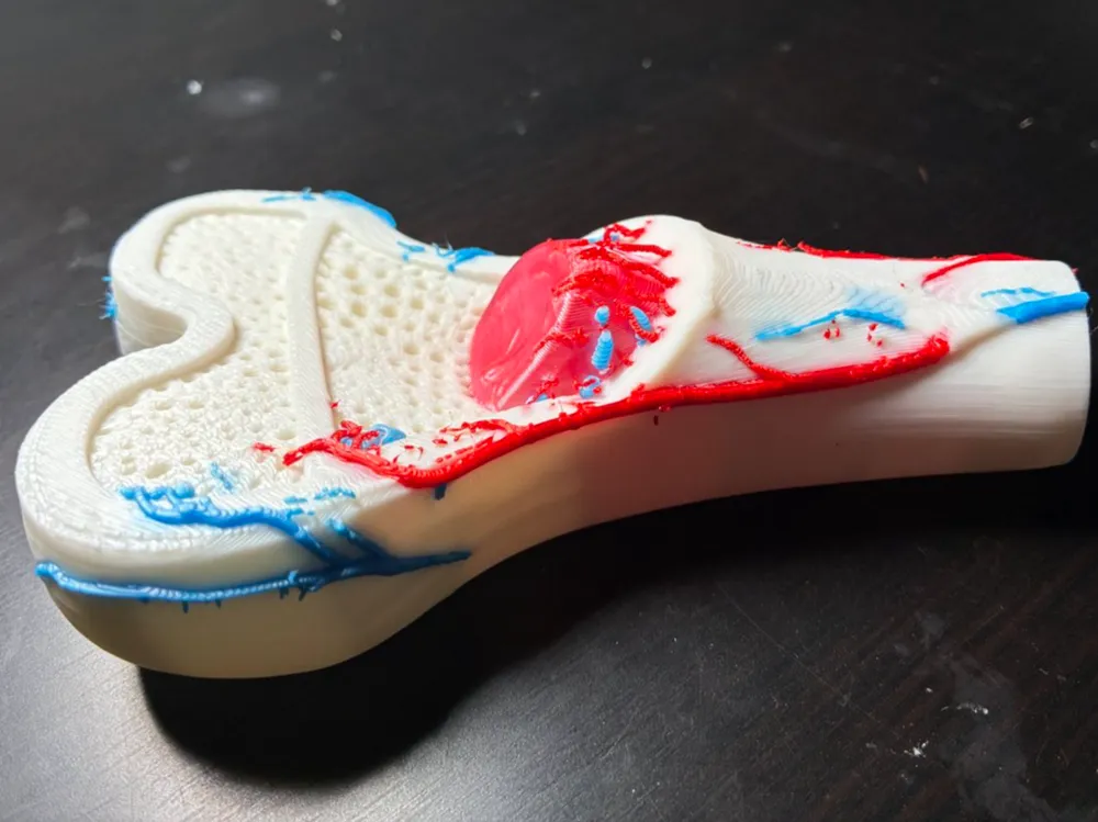
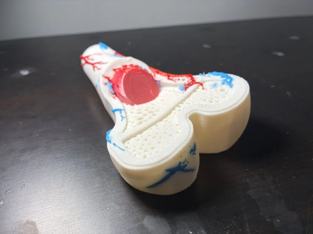
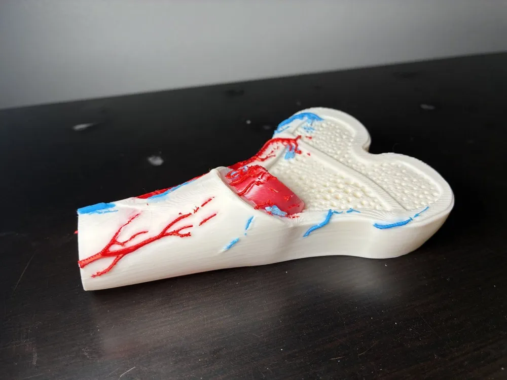
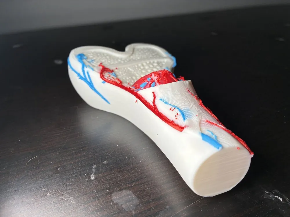
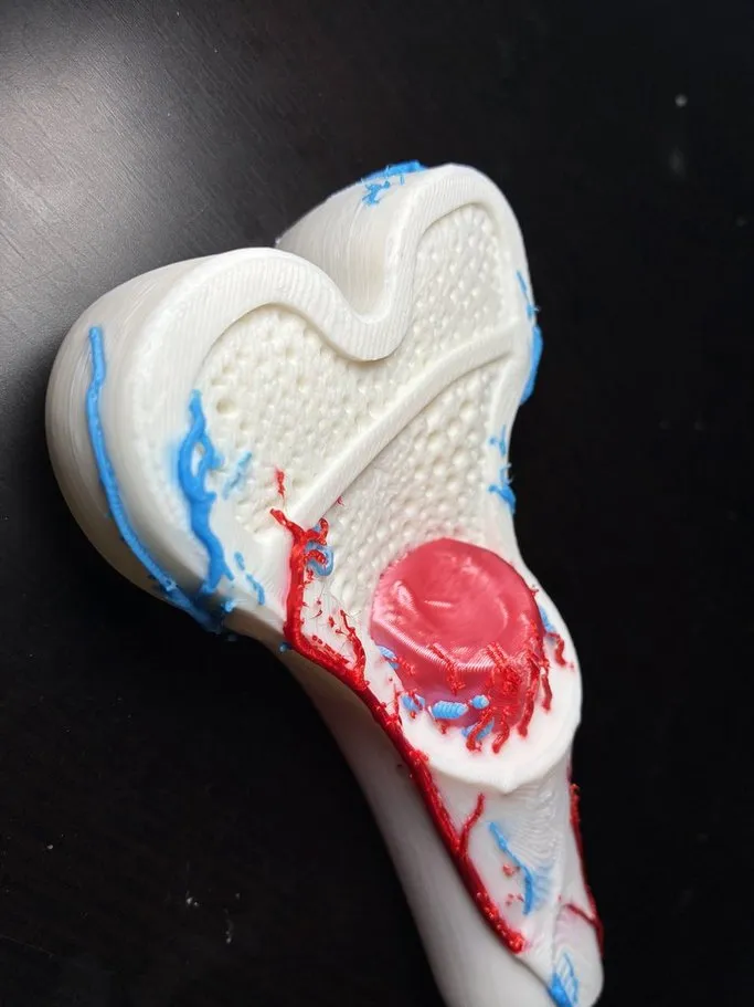
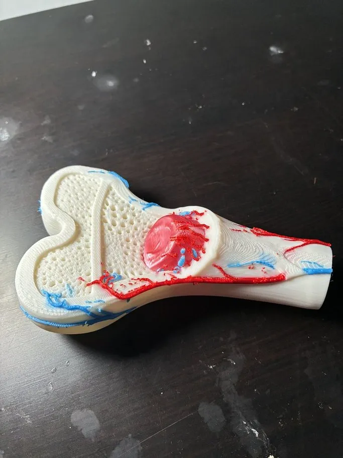
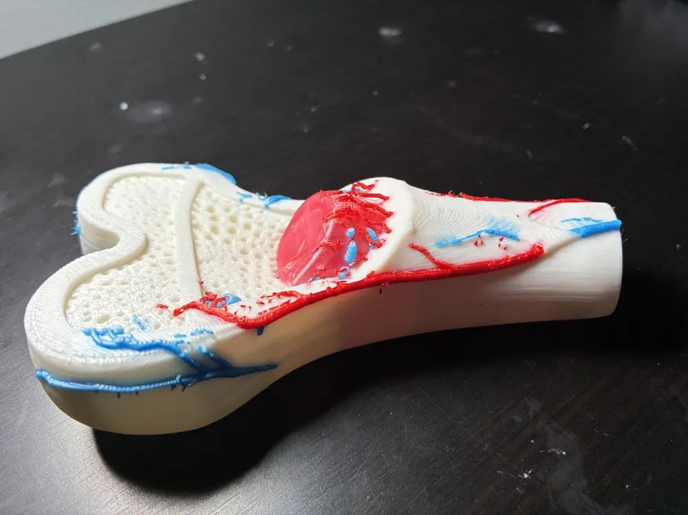
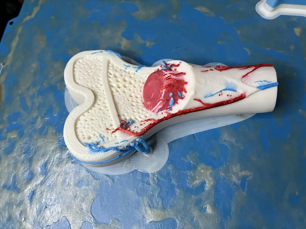
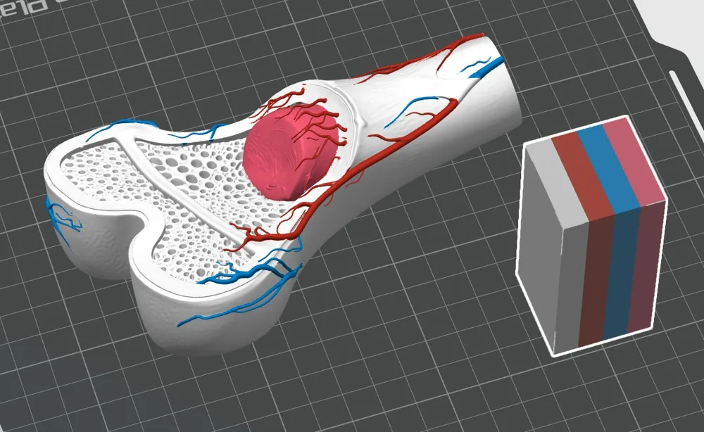
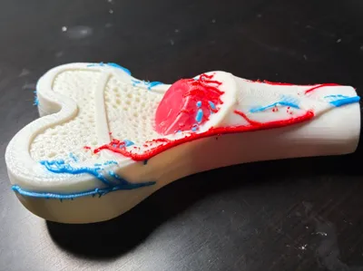
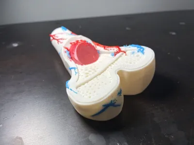
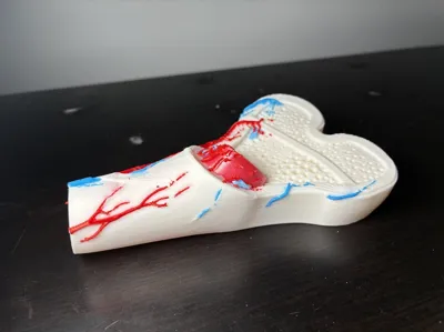
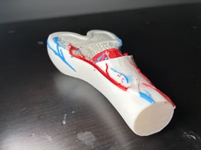
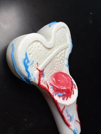
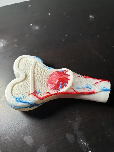
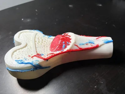
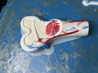
Dive deep into human biology with this high-fidelity, multi-color educational model of a long bone (Femur/Humerus cross-section). This model is designed specifically for students, medical professionals, and science enthusiasts to visualize what lies beneath the "hard" surface of our skeletal system.
The design features a strategic cutaway to reveal the complex layers of living tissue. It is an indispensable tool for classrooms or home-schooling environments where a 2D textbook image simply isn't enough to convey the 3D reality of biological architecture.
Key Anatomical Features Included:
- Compact Bone: The dense, hard outer layer that provides strength.
- Spongy Bone (Cancellous): The intricate, porous lattice structure shown in the epiphysis.
- Bone Marrow: Represented by the central red core, the powerhouse of blood cell production.
- Epiphyseal Line: The visible "growth plate" remnant where the bone once lengthened.
- Blood Vessels: Detailed arterial and venous pathways (Red and Blue) showing how bone is a living, breathing organ.
Print Settings & Material Recommendations
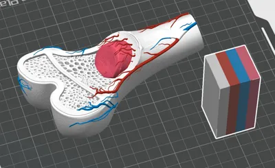
To achieve the vibrant, anatomical accuracy seen in the photos, this model is optimized for multi-color printing (AMS/Unitree).
Filament Recommendation: For the best results, I highly recommend using SUNLU Filament. Their tolerances are incredible for detailed anatomical prints like this.
- Bone Structure: SUNLU PLA Plus White – provides the perfect "bone" finish with superior strength.
- Marrow & Vessels: SUNLU PLA Meta Red and SUNLU PLA Meta Blue. The Meta series flows beautifully for those tiny vascular details.
Pro Tip for Multi-Color: Since this is a color-intensive print, check out my tutorial: How to remesh and color prints effectively in Bambu Studio and/or Snapmaker U1 to minimize waste and maximize detail!
History & Background
This model was developed out of a need for affordable, tactile medical training aids. Traditional medical-grade bone models can cost hundreds of dollars. By 3D printing this design, educators can provide every student with a hands-on experience, allowing them to trace the path of blood vessels and feel the difference between the porous spongy bone and the solid compact bone.
Remix Ideas & Expanded Use
This model is a perfect base for several other projects:
- Pathology Remix: Modify the marrow or spongy bone area to show the effects of Osteoporosis or Leukemia for clinical demonstrations.
- LED Diorama: Scale up and hollow out the vascular channels to run fiber-optic "glowing" blood vessels.
- Desk Organizer: Remix the flat end of the bone to hold pens or medical tools for a doctor’s office.
Keywords: Biology, Medicine, Skeleton, Anatomy, Education, STEM, Science Project, Doctor, Long Bone, Histology, Physiology.
#STEM #Anatomy #MedicalPrint #SUNLU #BambuLab #EducationalModel #Science
And if you've come this far, maybe you're willing to come a little further... - Andy Dufresne #TeachAManToFish
If you want to learn to do what I do (and stay out of my inbox so you can make your own changes to this file), click this link: https://youtu.be/iFfUVc33sTk
License

You shall not share, sub-license, sell, rent, host, transfer, or distribute in any way the digital or 3D printed versions of this object, nor any other derivative work of this object in its digital or physical format (including - but not limited to - remixes of this object, and hosting on other digital platforms). The objects may not be used without permission in any way whatsoever in which you charge money, or collect fees.


Comment & Rating (2)