


Print Profile(1)
Description
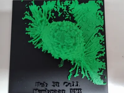
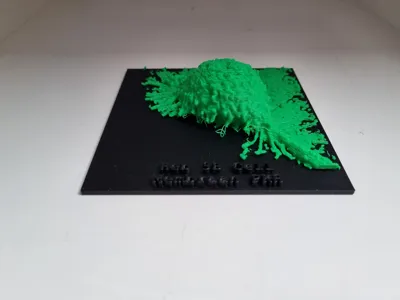
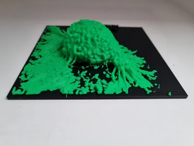
My first 3D model with a few day experience in 3D printing, so it might have some flaws. It was printed with a AMS. The original image is taken with a confocal laser scanning microscope. This gives a stack of 2D images that I rendered in ImageJ from which exported to a .stl file. This file was a bit complicated for Bambu studio so I simplified the model. After this I added a black backgroung because this was a fluorescent image, so the background is supposed to be black. It is a Hep3B cell a human epithelial cell line derived from a 8-year-old male with hepatocellular carcinoma, so it is tumor cell line. We stained the membrane with PKH67 a green fluorescent dye. This cell is going to a cell division which can be seen by the fact that is loosening from the coverslip on which it was cultured.
Comment & Rating (0)