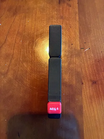
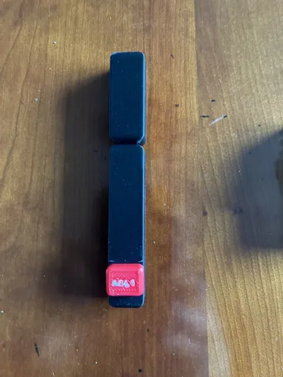
BCR-ABL FISH Philadelphia chromosome model


Print Profile(1)

Description
This model is used to demonstrate the detection of the BCR-ABL1 (t(9;22)(q34.1;q11.2) translocation ) fusion forming the Philadelphia Chromosome using FISH (Fluorescence In Situ Hybridisation) probes. This test is commonly used to identify this aberration in CML (Chronic Myelogenous Leukaemia) and some AML (Acute Lymphoblastic Leukaemia) cases.

Attribution for the diagram: AleksMjen, CC BY-SA 4.0 , via Wikimedia Commons
Information on FISH is available on wikipedia essentially the probes bind to areas of chromosomes and can be seen using a fluorescent microscope; seeing where and how the signals move gives information about what has happened to the region of the DNA to which the probe has bound.
The following picture shows what a BCR-ABL1 FISH probes looks like in use and is taken from Wikimedia Commons
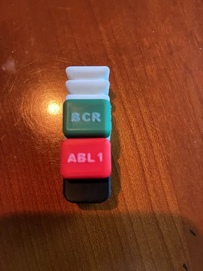
The 2 green and 2 red signals show BCR and ABL markers, the red and green signals together in the top left are showing a BCR-ABL rearrangement on a Philadelphia chromosome. These models can be used to demonstrate this

Magnets are embedded in the chromosomes and FISH probe markers so that the ABL1 tagged section of chromosome 9 can be swapped with the BCR tagged section of chromosome 22 giving a Philadelphia chromosome with the FISH markers indicating a BCR-ABL1 fusion (note this can appear as yellow when red and green combine). This is a reciprocal translocation where fragments of chromosome 9 and 22 swap place.
Magnets
Pauses are inserted in the file to allow for magnets to be inserted, make sure that you think about the orientation of the magnet, you want the two halves of each chromosome to attract and also they must attract the companion part for the Philadelphia chromosome. The FISH probes must also be placed correctly. ABL1 to the bottom section of 9 and BCR to the top of 22, it is easier to do this when you print the chromosomes first to check as you print.
Magnets for the chromosomes are 12mm dia by 3mm thick
Magnets for the FISH probes are 5mm dia by 1.5mm thick
I used the following magnets:
Chromosome magnets 12x3
If you leave the magnets out you can of course just sit the chromosome parts together. If you have a use for these please do let me know I'd love to hear what they are getting used for
Attribution for FISH photo:
No machine-readable author provided. Pmx assumed (based on copyright claims).,CC BY-SA 3.0 via Wikimedia Commons
License
You shall not share, sub-license, sell, rent, host, transfer, or distribute in any way the digital or 3D printed versions of this object, nor any other derivative work of this object in its digital or physical format (including - but not limited to - remixes of this object, and hosting on other digital platforms). The objects may not be used without permission in any way whatsoever in which you charge money, or collect fees.
Comment & Rating (0)