IV Ultrasound Trainer and Seek and Find


Print Profile(1)

Description
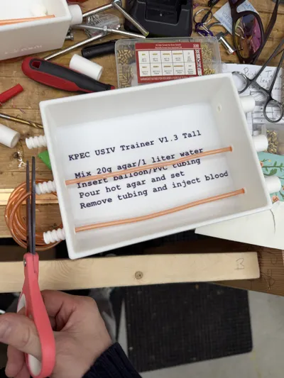
Ultrasound IV Training Model
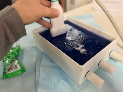
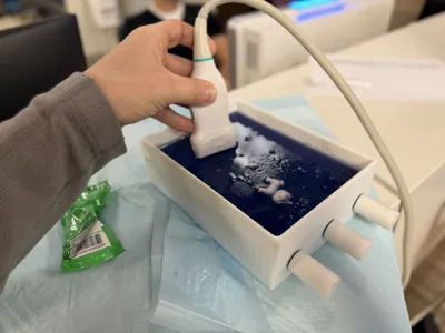
This model is designed as a realistic, reusable ultrasound IV access trainer. The cavity is designed to accommodate agar-based gel, a latex balloon “vessel,” and PVC tubing to form a compressible, ultrasound-visible target. It also includes several small structures to act as a “seek-and-find” challenge, helping users develop 3-dimensional orientation and confidence with ultrasound imaging.
Primary Function: Ultrasound IV Simulation
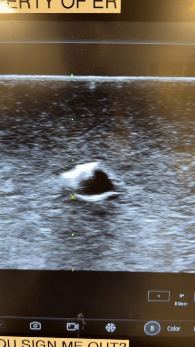
The central lumen of the model is sized for a standard balloon vessel. When filled with fluid, the balloon behaves like a compressible vein under ultrasound, providing a realistic practice target for cannulation.
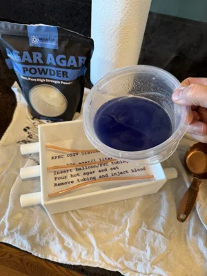
Agar Gel Instructions
Agar serves as the tissue-mimicking medium.
To mix the agar:
- Weigh out your water (for example 1149 g of water = 1149 mL).
- Use 2% agar by weight (e.g., 23 g agar powder for 1149 g water).
- Add agar powder to cold water and mix thoroughly.
- Slowly heat the mixture, stirring continuously.
- Bring it just to a boil, then remove from heat.
- Let the mixture cool slightly to reduce steam bubbles.
- WARNING: FOOD DYE MAY STAIN YOUR PROBE!!!!!
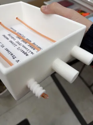
Balloon + Tubing Setup
- Inside the cavity, run a short segment of PVC tubing into the balloon to act as the vessel inlet.
- Position the tubing and balloon in the mold before you pour the agar.
- Pour agar around the balloon assembly and allow it to set completely.
Removing the Tubing
Once the agar has fully solidified:
- Gently twist and pull the PVC tubing free, leaving the balloon embedded in place.
- Use steady traction to avoid tearing the agar or the balloon.
- Fill a syringe with your preferred “blood” mixture (water + red dye works well).
- Remove all air from the syringe before injecting — air creates ultrasound artifacts.
- Inject into the balloon until it is full and compressible, mimicking a peripheral vein. Burp air as you fill.
Seek-and-Find Elements
Included in the print file are small, intentionally placed objects—bricks, paper clips, horse shoe, etc.
These serve two purposes:
- Improve 3D ultrasound orientation, especially recognizing depth, shadowing, and off-axis structures.
- Force the learner to sweep, fan, and track objects, building fine-motor control and probe awareness.
The extra objects turn this into a combined IV trainer and ultrasound exploration model, ideal for teaching residents, EMS, and nursing staff how to read complex images rather than relying on static patterns. It may take some time to perfect multiple pours.
Future Direction: I would like to create a cap that has a syringe-style injector to burp and refill veins.
Comment & Rating (7)